Melissa
Kaplan's
Chronic Neuroimmune Diseases
Information on CFS, FM, MCS, Lyme Disease, Thyroid, and more...
Last updated
January 1, 2014
|
Melissa
Kaplan's |
Fibromyalgia Pain Isn't All In Patient's Heads, New Brain Study Finds
fMRI scans give first objective measure of mysterious ailment, provide roadmap for future study
University of Michigan Health Services, June 2002
|
ANN ARBOR, MI - A new brain-scan study confirms scientifically what fibromyalgia patients have been telling a skeptical medical community for years: They're really in pain.
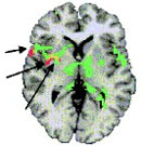
The results, published in the May issue of Arthritis & Rheumatism, the journal of the American College of Rheumatology, may offer the proof of fibromyalgia's physical roots that many doubtful physicians have sought. It may also open doors for further research on the still-unknown causes of the disease, which affects more than 2 percent of Americans, mainly women. Lead authors Richard Gracely, Ph.D., and Daniel Clauw, M.D., did the study at Georgetown University Medical Center and the National Institutes of Health, but are now continuing the work at the University of Michigan Health System. In an editorial in the same issue, Clauw and U-M rheumatologist Leslie Crofford, M.D., stress the importance of fibromyalgia research and care. To correlate subjective pain sensation with objective views of brain signals, the researchers used a super-fast form of MRI brain imaging, called functional MRI or fMRI, on 16 fibromyalgia patients and 16 people without the disease. As a result, they say, the study offers the first objective method for corroborating what fibromyalgia patients report they feel, and what's going on in their brains at the precise moment they feel it. And, it gives researchers a road map of the areas of the brain that are most - and least - active when patients feel pain. "The fMRI technology gave us a unique opportunity to look at the neurobiology underlying tenderness, which is a hallmark of fibromyalgia," says Clauw. "These results, combined with other work done by our group and others, have convinced us that some pathologic process is making these patients more sensitive. For some reason, still unknown, there's a neurobiological amplification of their pain signals." Further results from the study were presented last year at the ACR annual meeting. The project will continue later this year at UMHS, joining other fMRI fibromyalgia research now under way. For decades, patients and physicians have built a case that fibromyalgia is a specific, diagnosable chronic disease, characterized by tenderness and stiffness all over the body as well as fatigue, headaches, gastrointestinal problems and depression. Many patients with the disease find it interferes with their work, family and personal life. Statistics show that far more women than men are affected, and that it occurs mostly during the childbearing years. The ACR released classification criteria for fibromyalgia in 1990, to help doctors diagnose it and rule out other chronic pain conditions. Clauw and Crofford's editorial looks at the current state of research, and calls for rheumatologists to take the lead in fibromyalgia care and science. But many skeptics have debated the very existence of fibromyalgia as a clearly distinct disorder, saying it seemed to be rooted more in psychological and social factors than in physical, biological causes. Their argument has been bolstered by the failure of research to find a clear cause, an effective treatment, or a non-subjective way of assessing patients. While the debate has raged, neuroscientists have begun to use brain scan technology to identify the areas of the normal human brain that become most active during pain. A few studies have even assessed the blood flow in those areas in fibromyalgia patients during baseline brain scans. The new study is the first to use both high-speed scanning and a painful stimulus. In the study, fibromyalgia patients and healthy control subjects had their brains scanned for more than 10 minutes while a small, piston-controlled device applied precisely calibrated, rapidly pulsing pressure to the base of their left thumbnail. The pressures were varied over time, using painful and non-painful levels that had been set for each patient prior to the scan. The study's design gave two opportunities to compare patients and controls: the pressure levels at which the pain rating given by patients and control subjects was the same, and the rating that the two different types of participants gave when the same level of pressure was applied. The researchers found that it only took a mild pressure to produce self-reported feelings of pain in the fibromyalgia patients, while the control subjects tolerated the same pressure with little pain. "In the patients, that same mild pressure also produced measurable brain responses in areas that process the sensation of pain," says Clauw. "But the same kind of brain responses weren't seen in control subjects until the pressure on their thumb was more than doubled." Though brain activity increased in many of the same areas in both patients and control subjects, there were striking differences too. Patients feeling pain from mild pressure had increased activity in 12 areas of their brains, while the control subjects feeling the same pressure had activation in only two areas. When the pressure on the control subjects' thumbs was increased, so did their pain rating and the number of brain areas activated. But only eight of the areas were the same as those in patients' brains. In all, the fibromyalgia patients' brains had both some areas that were activated in them but not in controls, and some areas that stayed "quiet" in them but became active in the brains of controls feeling the same level of pain. This response suggests that patients have enhanced response to pain in some brain regions, and a diminished response in others, Clauw says. The study was supported in part by the National Fibromyalgia Research Association, the U.S. Army and the NIH. In addition to Clauw and Gracely, the research team included Frank Petzke, M.D.; and Julie M. Wolf, BA.
Special notes on this release If you have been diagnosed with fibromyalgia and would like to be contacted with further information about participating in fibromyalgia research at the University of Michigan, please call 800-742-2300, and enter category 6501. After listening to the recording, press 1 to leave your name, phone number, location and age after the tone. If you are seeking treatment for fibromyalgia, please call the Arthritis Foundation at 1-800-283-7800 to learn how to find a specialist near you. You can also visit their web site, www.arthritis.org, and enter your ZIP code to find the chapter nearest you. Functional Magnetic Resonance Imaging Evidence of Augmented Pain Processing in Fibromalgia [full text from UMich]
|
http://www.anapsid.org/cnd/diagnosis/brainpain.html
© 1994-2014 Melissa Kaplan or as otherwise noted by other authors of articles on this site
![]()